Science - Class 9

Topic outline
-
NCERT Solutions for Class 9 Science - Tissues (Biology), NCERT Textbook Solutions for Class 9 Science, NCERT Solutions For Class 9 Biology, Tissues - Class 9th NCERT Solutions Science, NCERT Solutions For Class 9 Biology Science Chapter 6 - Tissues, Science - Biology - Class 9 (CBSE/NCERT) - Chapter 6 – Tissues – Questions and Answers/Notes/Worksheets, CBSE Class 9 - Biology – Chapter 6 - Tissues Practice Pages, Extra Question and Answer based on NCERT for Class 9th, Science Biology, CBSE Grade IX free Worksheets PDF Tissues exemplar question answer, NCERT Book question answer, Science Question bank on Tissues for ninth standard, Differentiate between sclerenchyma and parenchyma tissues. Draw well labelled diagram. List the characteristics of cork. How are they formed? Mention their role. (a) Differentiate between meristematic and permanent tissues in plants (b) Define the process of differentiation. Name any two simple and two complex permanent tissues in plants. Describe the structure and function of different types of epithelial tissues. Draw diagram of each type of epithelial tissue.
-
Tissues
Q72. Differentiate between sclerenchyma and parenchyma tissues. Draw well labelled diagram.
Ans.
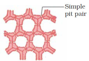
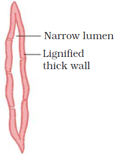
Sclerenchyma
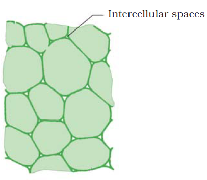
Parenchyma
1. It is a dead tissue.
1. It is a living tissue.
2. The cells are thick walled.
2. The cells are thin walled.
3. Cell cavity is narrow.
3. Cell cavity is wide.
4. It provides mechanical strength.
4. It stores nutrients and water especially in the stems and roots.
5. Intercellular spaces are absent in between the cells.
5. Intercellular spaces are abundant so that the tissue cells are loosely packed.
6. Transverse section
Longitudinal section
6. Transverse section
Longitudinal section
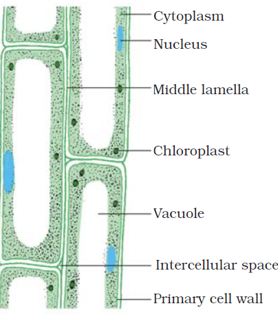
Image From NCERT
Q73. List the characteristics of cork. How are they formed? Mention their role.
Ans. Characteristics of cork
1. Cork is the outer protective tissue of older stems and roots.
2. The mature cork cells become dead and filled with tannins, resins and air.
3. Cork is a compact tissue.
4. Cork consists of several layers of cells.
5. Cork cells are impermeable due to deposition of suberin in their walls.
Formation of cork
As plants grow older, the outer protective tissue undergoes certain changes. A strip of secondary meristem replaces the epidermis of the stem. Cells on the outside are cut off from this layer. This forms the several-layer thick cork or the bark of the tree. Cells of cork are dead and compactly arranged without intercellular spaces. They also have a chemical called suberin in their walls that makes them impervious to gases and water.
Functions of cork
Cork is protective in function. Cork cells prevent loss of water from plant body. It prevents infection and mechanical injury. Cork is light and does not catch fire easily. Due to these properties, cork is used as insulators, shock-absorbers, linoleum, and sports goods.
Q74. Why are xylem and phloem called complex tissues? How are they different from one other?
Ans. Complex tissues are made of more than one type of cells. All these cells coordinate to perform a common function. Xylem and phloem are examples of such complex tissues. They are both conducting tissues and constitute a vascular bundle.
Difference between xylem and phloem
Xylem
Phloem
1. Xylem consists of tracheids, vessels, xylem parenchyma and xylem fibres.
1. Phloem is made up of four types of elements: sieve tubes, companion cells, phloem fibres and the phloem parenchyma
2. It transports water and minerals.
2. It transports food materials.
3. Conduction is mostly unidirectional. (from roots to apical parts of the plant)
3. Conduction is bidirectional. (from leaves to storage organs or growing parts ; or from storage organs to growing parts of plants)
4. Conducting channels are tracheids and vessels.
4. Conducting channels are sieve tubes.
5. Most of the components of xylem except xylem parenchyma are dead cells.
5. Most of the components of phloem except phloem fibres are living cells.
Q75. (a) Differentiate between meristematic and permanent tissues in plants (b) Define the process of differentiation. Name any two simple and two complex permanent tissues in plants.
Ans. (a) Difference between meristematic and permanent tissues
Meristematic tissues
Permanent tissues
1. Its component cells are living, small, spherical or polygonal and un-differentiated.
1. Its components cells may be living or dead. They are large, differentiated with different shapes.
2. The cytoplasm is dense and vacuoles are nearly absent as they are metabolically active.
2. Large central vacuole occurs in living permanent cells as, they are less metabolically active.
3. The cell wall of its cells is thin and elastic.
3. The cell wall of its cells may either thin or thick.
4. The nucleus of each cell of this tissue is large and prominent.
4. The nucleus is less conspicuous.
5. It is a simple tissue.
5. It can be simple, complex or specialized.
6. Cell organelles of its cells are simple.
6. Cell organelles of its cells are well developed.
7. Its cells grow and divide regularly.
7. Its cells do not normally divide.
8. It provides growth to the plant.
8. It provides protection. Support, conduction photosynthesis, storage, etc.
(b) The process of taking up a permanent shape, size, and a function is called differentiation.
(c) Simple permanent tissues in plants are parenchyma, collenchyma and sclerenchyma. Complex permanent tissues in plants are xylem and phloem.
Q76. Describe the structure and function of different types of epithelial tissues. Draw diagram of each type of epithelial tissue.
Ans. Types of epithelial tissue:
Simple squamous epithelium - In cells lining blood vessels or lung alveoli, where transportation of substances occurs through a selectively permeable surface, there is a simple flat kind of epithelium. This is called the simple squamous epithelium. Simple squamous epithelial cells are extremely thin and flat and form a delicate lining. The oesophagus and the lining of the mouth are also covered with squamous epithelium. The skin, which protects the body, is also made of squamous epithelium.
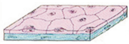
Image From NCERT
Stratified squamous epithelium - Skin epithelial cells are arranged in many layers to prevent wear and tear. Since they are arranged in a pattern of layers, the epithelium is called stratified squamous epithelium.
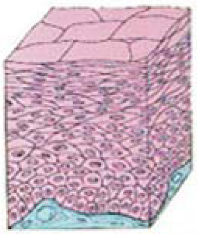
Image From NCERT
Ciliated columnar epithelium - Where absorption and secretion occur, as in the inner lining of the intestine, tall epithelial cells are present. This columnar (meaning ‘pillar-like’) epithelium facilitates movement across the epithelial barrier. In the respiratory tract, the columnar epithelial tissue also has cilia, which are hair-like projections on the outer surfaces of epithelial cells. These cilia can move, and their movement pushes the mucus forward to clear it. This type of epithelium is thus ciliated columnar epithelium.
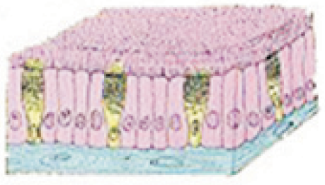
Image from NCERT
Cuboidal epithelium - Cuboidal epithelium (with cube-shaped cells) forms the lining of kidney tubules and ducts of salivary glands, where it provides mechanical support. Epithelial cells often acquire additional specialisation as gland cells, which can secrete substances at the epithelial surface. Sometimes a portion of the epithelial tissue folds inward, and a multicellular gland is formed. This is glandular epithelium.
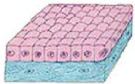
Image from NCERT
-